Insect based feed for animals
in aquaculture 
in the EU

25.10.2017  /  Scienceandmore  /  Category: Animal Biology

Recent changes in EU legislation allow feeding of animals in aquaculture with processed animal protein (PAP) from insects since July 2017. This amendment mainly affects insect meal (dried insects ground to meal) that consists of protein to a big proportion, especially when it is defatted. Oil from insects, as well as fresh or unprocessed dried insects are not included in this restrictions by EU legislation and can be fed to animals in aquaculture, pets, poultry and pigs. Here the question arises, why would insect meal be preferred over fresh and dried insects? Insect meal can be mixed with other feed components such as ground grains and soy to form a mixture of desired composition that is then pressed in pellets for better and more convenient feeding to animals.

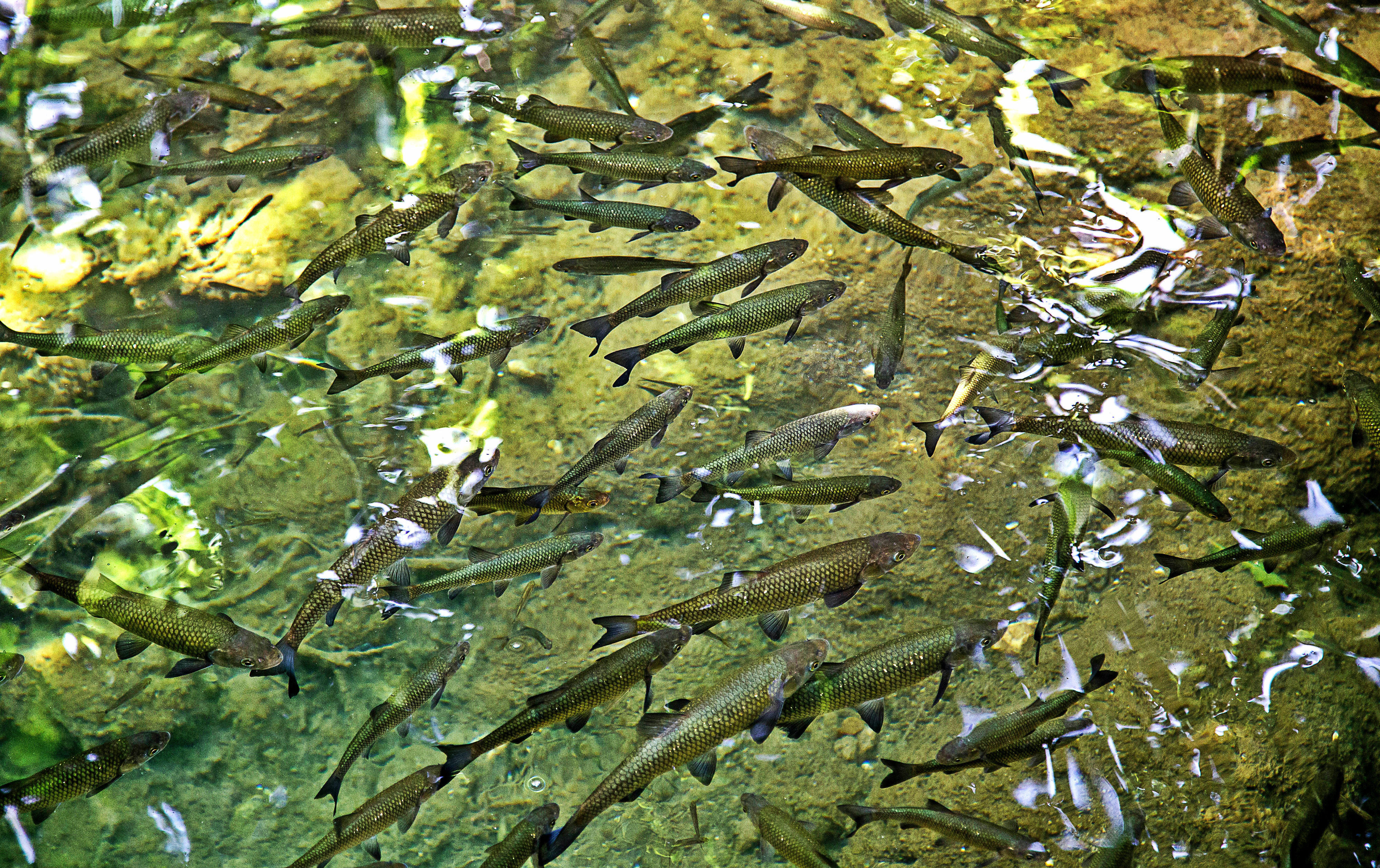
Animals in aquaculture are mostly reared by the five EU member states Spain, UK, France, Italy and Greece, who produce around 75 % of the animals in the EU. 294,000 tons of animals in aquaculture were reared by Spain (equals 23.0 % of the EU total, with a worth of approximately €513 million), 212,000 tons by the UK (16.6 %, €995 million), 180,000 tons by France (14.1 %, €627 million), 149,000 tons by Italy (11.6 %, €437 million) and 106,000 tons by Greece (8.3 %, €463 million) (1). Here, the most relevant and important animals are molluscs and fish, and the following 10 species account for around 90 % of the total weight in the EU in descending order:

1) Mediterranean mussel (Mytilus galloprovincialis), 2) Atlantic salmon (Salmo salar), 3) rainbow trout (Oncorhynchus mykiss), 4) blue mussel (Mytilus edulis), 5) gilt-head seabream (Sparus aurata), 6) Pacific oyster (Magallana gigas), 7) European bass (Dicentrarchus labrax), 8) common carp (Cyprinus carpio), 9) Japanese carpet shell (Venerupis philippinarum), and 10) Atlantic bluefin tuna (Thunnus thynnus).

This is stated with the annotation that the EU consists of different countries with different geography, and that the various fish species also have different natural habitats. 95% of the Atlantic salmon is reared in the UK, and 87% of Pacific oyster in France for instance (1). At the same time, other species have wider dissemination. Rainbow trout for example is reared by 17 of the EU member states and the common carp by 13 states as one of their major fish species (2).

salmon

In terms of species-specific feed in the wild, the three molluscs Mediterranean mussel, blue mussel and Pacific oyster consume bacteria, phytoplankton and other small organic matter. In aquaculture, their cultivation does not require additional feed (3-5).

The stated seven fish species are carnivorous or omnivorous and a major part of their diet in the wild are other fish and shellfish, but also insects (6-9).  As of yet, fish in aquaculture have been fed with fishmeal and plant-based feeds, as well as oil from fish and plants. Due to the increasing prices of fishmeal and fish oil, more and more plant-based material from legumes, oil seeds or cereal gluten has been introduced to animal feed. It seems, however, difficult to substitute large amounts of fishmeal and especially fish oil with plant based material. They have disadvantages compared to fishmeal, such as less palatability, anti-nutritional components, high fibre and non-starch polysaccharides content, and a fatty acid and amino acid profile that were proposed to be less suitable than the ones of fishmeal (1).

Insect meal could be a beneficial addition in fish feed and could substitute parts of fishmeal and plant content, since the amino acid profile of insects is more comparable to fishmeal than the one of plants to fishmeal (11). However, depending on the utilised insect species, insect proteins could still be low in the amino acids such as methionine and lysine that potentially would make it necessary to supplement these in animal feeds, specifically for growing animals. This also suggests that insect meal could only partially substitute fishmeal (12,13). The combination of different insect species could compensate the lack of certain amino acids in one of the species. Considering the amino acid profiles of black soldier fly and house fly larvae, they partly complement each other and together they match the profile of fishmeal much closer than the individual ones. The low levels of tryptophan in black soldier fly larvae could be compensated by the higher amounts in house fly larvae for instance.

Fish oil contains higher amounts of omega-3 fatty acids than vegetable oil that are in particular important for carnivorous species such as salmon and trout. The amount of these fatty acids could be insufficient for the fish if the plant content of their feed would be higher (10). The ratio of alpha-linolenic acid (one of the three omega-3 fatty acids) to linoleic acid in insect oils is not as good as in fish oils, but in general better than in vegetable oils. Nevertheless, this is one of the limiting factors for the utilisation of insect meal and insect oil in fish feed, since fish need these fatty acids due to limited metabolic capabilities to synthesis them or due to better growth and development when these fatty acids are provided in their diet (13). However, investigations showed that the fatty acid composition of insects can be modified by modulating the content of their substrate (12,13), and thus potentially adjusted to fit more closely the requirements of a specific fish species.

Fish diets contain relatively low levels of carbohydrates, and the most common carbohydrate would be chitin that is probably digested to a degree by fish. Investigations showed chitinase activity, an enzyme that is involved in chitin digestion, in several fish species. The addition of chitin to fish feed of marine fish has even been suggested to have beneficial effects on these fish. Insects contain low levels of carbohydrates that were estimated to be less than 20 %, and chitin forms the majority of it. However, it is agreed upon that chitin is another limiting factor for the utilisation of insect in fish feed (2014).

Insects are in general relatively low in calcium compared to fish meal, although there are exceptions like the black soldier fly that is high in calcium. Soy is also characterised by low levels of calcium. However, similar to the fatty acid content, calcium content of insects can be affected by the calcium content of their substrate. Nevertheless, addition of calcium could be necessary to ensure optimal development and growth of fish. Phosphorus levels are also relatively low in insects, as well as in soy, compared to fishmeal. An important factor for fish feed is the ratio of calcium to phosphorous and would possibly need adjustment to reach 1.1 to 1.4 as found in fishmeal (13).

According to EU legislation, seven insect species are allowed for the production of animal feed in aquaculture (Regulation (EU) N° 2017/893):

1) black soldier fly (Hermetia illucens), 2) housefly (Musca domestica), 3) mealworm or yellow mealworm (Tenebrio molitor), 4) lesser mealworm or litter beetle (Alphitobius diaperinus), 5) house cricket (Acheta domesticus), 6) tropical house cricket or banded cricket (Gryllodes sigillatus), and 7) Jamaican field cricket (Gryllus assimilis).

These species are regarded as non-pathogenic and do not pose a risk for human, animal or plant health. Furthermore, they do not transmit human, animal or plant pathogens, i.e. are not regarded as vectors, are not protected species or invasive alien species. In fact, they are now classified as „farm insects” and have a similar status as livestock (according to EG 1069/2009 of the new legislation). The new legal status of these seven insects has certain additional consequences and regulation that need to be abided by. For animal feed production purpose, a cultivation of these insects is not allowed on substrate that contains kitchen or food scraps, meat or bone meal, liquid manure or faeces (according to EG 999/2001 and EG 767/2009).


Allowed are substrates of:

1) non-animal origin or

2) animal origin that fall within category 3 substances, specifically fish meal, blood products of non-ruminants, Dicalciumphosphate and Tricalciumphosphate, hydrolysed proteins from non-ruminants, hydrolysed proteins of ruminants and their hides, gelatine and collagen of non-ruminants, eggs and egg products, milk and dairy-based substances, colostrum, honey, and rendered fats (amended schedule XIV, chapter I, clause 2, section 5 b and c of the EU 142/2011).

Significantly, high importance was placed on the exclusion of unprocessed products of ruminant origin to prevent potential cross-contamination with prions. Prions were attributed for the catastrophic outbreak of BSE in the 90s. These substrate regulations also apply for insect meal that is imported from non-EU counties into EU countries.


Next article: Black soldier fly larvae as fish feed in the EU

References

1. Eurostat (2016) Dive into aquaculture in the EU. [online] Eurostat. Available at: http://ec.europa.eu/eurostat/en/web/products-eurostat-news/-/DDN-20171018-1 [Accessed 19.10.2017].
2. Eurostat and Eumofa (2013) European Commission – Fisheries – 4. Fisheries and aquaculture production [online]. Available at: https://ec.europa.eu/fisheries/4-fisheries-and-aquaculture-production_en [Accessed 20.10.2017].
3. MarLIN (2006) BIOTIC – Biological Traits Information Catalogue. [online] Marine Life Information Network. Plymouth: Marine Biological Association of the United Kingdom. Available at: www.marlin.ac.uk/biotic [Accessed 19.10.2017].
4. Department of Agriculture, Forestry and Fisheries (DAFF) (2017) Mediterranean mussel Mytilus galloprovincialis. [online] Available at: http://www.nda.agric.za/doaDev/sideMenu/fisheries/03_areasofwork/Aquaculture/BIODIVERSITY/M.%20galloprovincialis%20BRBA%2012.12.12.pdf [Accessed 19.10.2017].
5. FAO (2017) Ruditapes philippinarum (Adams & Reeve, 1850). [online] Cultured Aquatic Species Information Programme. Available at: http://www.fao.org/fishery/culturedspecies/Ruditapes_philippinarum/en [Accessed 25.10.2017].
6. Rochard, E. and P. Elie (1994) La macrofaune aquatique de l’estuaire de la Gironde. Contribution au livre blanc de l’Agence de l’Eau Adour Garonne. In État des connaissances sur l’estuaire de la Gironde (ed. by Mauvais, J.-L. and Guillaud, J.-F.), pp 1-56. Agence de l’Eau Adour-Garonne, Éditions Bergeret, Bordeaux, France.
7. Frimodt, C. (1995) Multilingual illustrated guide to the world’s commercial warmwater fish. Fishing News Books, Osney Mead, Oxford, England.
8. Kottelat, M. and Freyhof, J. (2007) Handbook of European freshwater fishes. Publications Kottelat, Cornol and Freyhof, Berlin.
9. ICCAT (2009) Report of the standing committee on research and statistics (SCRS). Madrid, Spain.
10. Scientific, Technical and Economic Committee for Fisheries (STECF) (2014) The Economic Performance of the EU Aquaculture Sector (STECF 14-18) (Ed. Nielsen, R., Motova, A.), pp. 403. Publications Office of the European Union, Luxembourg, EUR XXXX EN, JRC XXXX, xxx pp.
11. PROteINSECT (2016) Insect Protein – Feed for the Future. [online] Minerva Communications UK Ltd. Available at: https://www.fera.co.uk/media/wysiwyg/our-science/proteinsect-whitepaper-2016.pdf [Accessed 19.10.2017].
12. Makkar, H.P.S., Tran, G., Heuzé, V., Ankers, P. (2014) State-of-the-art on use of insects as animal feed. Animal Feed Science and Technology, 197, 1-33.
13. Tran, G., Heuzé, V., Makkar, H.P.S. (2015) Insects in fish diets. Animal Frontiers. 5(2):37-44.

Insects – the new animal feed in the EU

24.10.2017  /  Scienceandmore  /  Category: Animal Biology

Could insects be used to feed livestock and what would be the benefits to use insects compared to conventional animal feed?

This is the first of several consecutive articles that discuss the utilisation of insects as animal feed. Here, the legislative aspects for insects in the EU, animal feed in general, the use of insects for certain animals, and potential hazards are introduced.

Introduction

Livestock are of paramount importance for human diet and even though the proportions vary from country to country, on average around 95 % of the worldwide population eats meat (1).

In 2016, livestock that mainly consists of cows, pigs, poultry and shellfish/fish has been fed with more than one billion tons of feed. Approximately 44 % of the total animal feed was produced for poultry, followed by around 27 % for pigs, 22 % for cattle, and 4 % for animals in aquaculture (2).

Poultry, which is a generic term for mostly chickens, turkeys, quails, ducks and geese, is mainly fed with grains but also soy and fishmeal as a protein source. Pigs as omnivorous animals are also fed with grains, soy and fishmeal, as well as with oilseed meals, root crops and legumes. Cattle as herbivorous animals get grass and small amounts of grain and soy. Fish in aquaculture are mostly carnivorous, such as salmon and trout, and are commonly fed fishmeal in pellet form, but also soy, grains and legumes as cheaper protein and nutrient sources (3-6).

It is apparent that fishmeal and soy are crucial parts of animal feed. Approximately 16 to 17 million tons of wild fish (caught in the ocean) are processed into fishmeal and fish oil (including 5 million tons of fish trimmings) annually. The majority of this fishmeal and oil is used in aquacultures to rear fish for human consumption (6). However, prices for fishmeal have been risen due to high demand, especially since 2010/11, and in consequence to increased production of fish in aquacultures, farmers started to rely more on protein-rich plant-based materials for feed (6-8). The crops that are used are mainly imported from non-EU countries. In fact, only around 30 % of these crops are produced inside the EU and 70 % are imported from Brazil, Argentina and the USA. The import amounts to approximately 30 million tons annually, and soy is a major part of it. Significantly, 80% of the world’s soybean production is used as animal feed (6,7).

soya-83087_960_720

An alternative to fishmeal and soy could have been found in insect meal. Insects in general consist of approximately 40 to 60 % protein and up to 36 % of fat that could be fed separately as insect meal and insect oil (9). Insects are naturally eaten by cattle, pigs, poultry and fish as part of their species-appropriate diet (7). It is also not a new idea to use insects as a part of animal feed, specifically insect protein. However, in Europe during the 90s, there was a wide-ranging mad cow disease (bovine spongiform encephalopathy (BSE)) occurrence that was caused by feeding cows proteins from the remains of other cows, i.e. processed animal protein. Subsequently, almost all processed animal protein including insect protein was prohibited to be used as animal feed in the EU. An exemption was protein from fish.

Nevertheless, efforts were made to change EU legislation and finally on 1st of July 2017, processed animal protein from insects was allowed to be used for animals in aquaculture (EU-enactment 2017/893) … however, only seven specific insect species received approval!

Before addressing the approved insect species, here are the most important arguments in favour of insects:

1) Insects meal can partly substitute for fishmeal and plant-based components in feed.  Studies in livestock showed that insect meal could substitute fishmeal in feed to a certain degree (9). Fishmeal that is used to feed poultry, pigs and fish in aquaculture is produced from wild fish caught in the oceans. Using insect meal instead could therefore possibly prevent overfishing.

Plant-based components in fish feed have disadvantages compared to fishmeal, such as less palatability, anti-nutritional components, high fibre content, non-starch polysaccharides, and a different amino acid profile. Insects, on the contrary, are much lower in fibre and anti-nutritional content, and it has been proposed that they have a better suited amino acid profile than plant based components (10,11).

Since insect meal is only allowed to feed animals in aquaculture in the EU, the following article will focus on the utilisation of insect meal in this sector.

2) Insects require less space and energy for cultivation compared to soy. Soybean cultivation in the South American countries led to a drastic change in land use, most significant to a deforestation, as well as decreased soil fertility, ruined biodiversity and the use of tremendous amounts of water (6). Even though soy will most likely remain as one of the protein source in animal feed in the near future, insect protein could partly replace it. Consequently, the impact of soy cultivation on the environment could be reduced.

Compared to soybeans, insects are very efficient in utilising energy. Since they are cold-blooded, they do not expend energy to regulate their body temperature, so energy can be used for growth and development. A study showed that the production of 1 ton of crickets, which equals to around 600 kg of protein, requires around 2.8 tons of feed and a surface of 3,100 m2. For soy, estimations suggest that the production of 1 ton, which equals to around 50 kg of protein, requires around 3,200 m2 of land and takes one year (9,12,13). The life cycle of the cricket species Acheta domesticus on the other hand takes only two to three months! Obviously, the cricket feed/substrate also needs to be calculated in order to estimate if rearing crickets would be feasible, ecologically and economically. But that is the great aspect of insect cultivation… insects can feed on organic waste.

3) Insects have a broad range of substrates they can thrive on, such as food waste. In Germany, around 9.6 million tons, in Italy 5.7 million tons, in the UK 5 million tons, in France 3.7 million tons, and in all 28 EU member countries combined 31.2 million tons food waste were produced in 2014. This comprises animal and vegetable waste (14). This food waste could be used to rear insects and represents a cheap and even revenue-generating substrate. After rearing, the residual substrate could be used as fertiliser for crop production or as material to remediate soil, as it is still rich and accessible in nutrients and minerals. Investigations showed that the larvae of black soldier flies, one of the insect species that is approved for animal feed production, reduced the volume of organic waste by up to 60 % in just 10 days (7).

However, EU legislation does not allow the utilisation of food waste as substrate for insects that are meant for animal feed production (according to EG 999/2001 and EG 767/2009), due to the danger of a possible accumulation of chemicals and toxins in insects. These potentially hazardous substances could eventually accumulate in animals, generate allergens or, even worse, disease and would pose harm to the animals and humans upon consumption of the livestock.

4) Insects can be cultivated all year around. Most crop plants used for animal feed production are primarily cultivated in the field and are therefore seasonally restricted in their cultivation. Insects, however, can be reared all year around indoors. They would however require stable temperatures for optimal growth and development.

grasshopper-2497947_960_720

5) The use of insects as animal feed could lead to more independence from the import of raw material for animal feed production and result in a more stable market for animal feed in the EU. This is especially important since the demand of Asian countries for raw material continuously increases. Additionally, the American countries that are the major producers of soybeans are not liable to the strict regulations of GMO use of EU legislation (6,7).

Sometimes, implementing changes to improve handling of environmental and ecological issues can be motivated by financial gains. In 2014, 980 million tons of animal feed were produced, and in 2016 the production increased to over 1 billion tons worldwide, which translate to a value of approximately $460 billion (approximately €390 billion) (15,2), and makes the animal feed sector a lucrative sector for innovations. These numbers represent the whole animal feed sector but the changes in EU legislation only affect the aquaculture sector. Here, the production of animals in aquaculture increases constantly. In 2012, it already reached 1.108 million tons of animals with a commercial value of almost €3.4 billion in the EU alone (7). It has to be pointed out that the EU is the no. 1 fish importer worldwide, with imports valued at US$54 billion (approximately €45.6 billion) in 2014 (16). Similarly, feed for domestic animals in aquaculture is mostly imported as well (only 5.9 million tons of feed for these animals was produced in Europe itself in 2014) (15). Production of sustainable feed and rearing of greater numbers of animals in aquaculture in the EU could decrease the need to import them to satisfy domestic consumption.

References

1. FOE (2014) Meat Atlas – Facts and Figures About the Animals We Eat. [online] FOE Europe. Available at: https://www.foeeurope.org/sites/default/files/publications/foee_hbf_meatatlas_jan2014.pdf [Accessed 16.10.2017].
2. Alltech (2016) Global Feed Survey. [online] Alltech. Available at: https://go.alltech.com/alltech-feed-survey [Accessed 16.10.2017].
3. Food Standards Agency. What farm animals eat. [online] Food Standards Agency. Available at: https://www.food.gov.uk/business-industry/farmingfood/animalfeed/what-farm-animals-eat [Accessed 16.10.2017].
4. Lázaro, R., Mateos, G.G., Latorre, M.M., Javier, P. (2015) Whole soybeans in diets for poultry. American Soybean Association
5. McGlone, J., Pond, W.G. (2003) Pig production: biological principles and applications. pp. 191. Cengage Learning, NY, USA.
6. Stamer, A. (2015). Insect proteins – a new source for animal feed: The use of insect larvae to recycle food waste in high-quality protein for livestock and aquaculture feeds is held back largely owing to regulatory hurdles. EMBO Reports, 16(6), 676–80.
7. PROteINSECT (2016) Insect Protein – Feed for the Future. [online] Minerva Communications UK Ltd. Available at: https://www.fera.co.uk/media/wysiwyg/our-science/proteinsect-whitepaper-2016.pdf [Accessed 16.10.2017].
8. Van Huis, A., van Itterbeeck, J., Klunder, H., Mertens, E., Halloran, A., Muir, G., Vantomme, P. (2013) Edible insects: future prospects for food and feed security. FAO Forestry Paper 171: 89-97.
9. Makkar, H.P.S., Tran, G., Heuzé, V., Ankers, P. (2014) State-of-the-art on use of insects as animal feed. Animal Feed Science and Technology, 197, 1-33.
10. Sanchez-Muros, M.-J., F.G. Barroso, and F. Manzano-Agugliaro (2014) Insect meal as renewable source of food for animal feeding: A review. J. Clean. Prod. 65:16–27.
11. Tran, G., Heuzé, V., Makkar, H.P.S. () Inserts in fish diets. Animal Frontiers 5(2): 37-44.
12. Langemeier, M., and E. Lunik (2015) International Benchmarks for Soybean Production. farmdoc daily (5):225.
13. Collavo, A., Glew, R.H., Huang, Y.S., Chuang, L.T., Bosse, R., Paoletti, M.G. (2005) House cricket small-scale farming. In: Ecological implications of minilivestock: potential of insects, rodents, frogs and snails. (ed. by Paoletti, M.G.), pp. 519–544. New Hampshire, Science Publishers.
14. Eurostat (2016) Waste generated by households by year and waste category – Animal and vegetable wastes. [online] Eurostat. Available at: http://ec.europa.eu/eurostat/tgm/refreshTableAction.do?tab=table&plugin=1&pcode=ten00110&language=en [Accessed 16.10.2017]
15. Alltech (2015) 2015 Global Feed Survey. [online] Alltech. Available at: https://www.alltech.com/sites/default/files/global-feed-survey-2015.pdf [Accessed 16.10.2017]
16. FAO (2016) The State of World Fisheries and Aquaculture 2016. Contributing to food security and nutrition for all. Rome.

Immune Response of Plants – Part 2

22.09.2017  /  Scienceandmore  /  Category: Plant Biology

This is the second part of the review about the plant immune response. For the first part, please click here.

Recently, the zig zag model for plant-pathogen interaction was criticised since it is based on the plant’s interaction with biotrophic microbes, i.e. pathogens that keep their host plans alive while feeding on them, and lacked integration of symbiotic interactions, or the response to necrotrophic pathogens. Hypersensitive response (HR), a form of programmed cell death, has been suggested as a major immune response of the plant in the zig zag model (1), and research suggests that it is effective against biotrophic microbes (2). In terms of an infection with necrotrophic pathogens, HR would, however, rather be a favourable result, since necrotrophic pathogens aim to kill plant cells in order to obtain nutrients. HR would therefore contribute to an invasion by necrotrophs and be counterproductive for the plant.

Also, the environmental context such as the plant’s previous encounters with pathogens, presence of beneficial microbes, etc. have not been considered in the zig zag model. In this model, inducible plant immune response is defined as a mechanism in which perception of MAMPs and microbial effectors result in immunity (PTI/ETI) or susceptibility (ETS). Instead, it was suggested that the plant immune response should rather be defined as a mechanism that integrates various signals and results in different outcomes which cannot simply be categorised into ETI, PTS and PTI (3). An alternative model to describe inducible plant immune response has been proposed by Cook et al. (4) with the invasion model. In this model, compounds that elicit the plant immune response are not categorised into the strict categories of MAMPs or effectors but into invasion patterns (IPs). These IPs are not defined from the point of perception and response by the host plant, but from the point of their main function in the microorganism or plant. IPs are for example microbial-derived compounds such as the fungal chitin and bacterial flagellin, which would have been described as MAMPs in the zig zag model, but also host plant-derived modified-self compounds that indicate an invasion. Modified-self compounds are for example damage associated molecular patterns (DAMPs) such as components of the exterior of the cell that are released after wounding. Well described DAMPs are pectin fragments that are released from the cell wall after penetration by a fungus for instance. However, the main function of pectin is in plant cell wall structure and not as indicator of invasion. DAMPs play an important role in plant immune responses but have not been integrated in the zig zag model (3).

Also, a classification of immune response eliciting compounds into MAMPs or effectors is objected to in the invasion model, since some of these compounds have been found to not fit into the categories of MAMPs and effectors (3).

Contrary to the zig zag model, in the invasion model the host plant’s responses are not classified into PTI or ETI, but into IP triggered responses (IPTRs). The difference is that perception of IPs (by invasion pattern receptors (IPRs)) results in a) continued symbiosis between invader and host plant, or b) end of symbiosis of invader and host plant; instead of immunity (PTI/ETI) or susceptibility (ETS) as conceptualised in the zig zag model. These outcomes of continuation or discontinuation of symbiosis are determined by the overall effect of elicited host plant responses (that can be synergistic and/or antagonistic) (4).

From the invader perspective, mechanisms that lead to a) the failure to supress IPTR, b) the suppression of IPTR (for example by biotrophic invaders), or c) the utilisation of IPTR (for example by necrotrophic invaders to drive cell death) determine if the symbiosis with the host plant is either continued or discontinued. Invaders may deploy IPs to influence the outcome of the symbiosis by manipulating IPTRs. When these IPs are used or the symbiosis is continued, further IPs could be released such as components that indicate a modification of host plant processes or DAMPs that in turn could be perceived by IPRs and lead to a discontinuation or continuation of the symbiosis, depending on the triggered plant responses (4).

In the zig zag model, plant immune response and resistance are associated with HR, which, however, would rather result in a susceptibility to necrotrophic pathogens instead of resistance. In the invasion model, infection by necrotrophs leads to the release of DAMPs such as pectin fragments from the cell wall, resulting in triggering of appropriate defence responses. Additionally, pro-death mechanisms utilised by necrotrophic microorganisms such as specific toxins to hijack the plants immune response to drive HR for instance did not really fit in the zig zag model but are integrated in the invasion model.

Symbiosis of host plants with beneficial microbes also depends on IPs (effectors) that suppress the plants defence response, similar to interactions with pathogens. Arbuscular mycorrhiza fungi, which are fungi that colonise the roots of a wide range of land plants and that are essential for the host plants development, growth and stress tolerance (5), also deploy effector to attenuate the plants immune response. This, however, does not represent susceptibility of the host plant (ETS in the zig zag model) in a narrow sense and, therefore, does not really fit into the zig zag model. In the invasion model on the other hand, arbuscular mycorrhiza fungi deploy IPs that suppress IPTRs, which otherwise would lead to a discontinuation of symbiosis (4).

Finally, it has to be pointed out that both the zig zag model and the invasion model are models after all that try to incorporate as many aspects of plant-microbe interaction and make sense of them. Nevertheless, models are generalisations and are therefore incomplete but form the basis for new scientific hypotheses and progress.

References

1. Jones, J.D. and Dangl, J.L. (2006) The plant immune system. Nature, 444, 323-9.

2. Coll, N.S., Epple, P., Dangl, J.L. (2011): Programmed cell death in the plant immune system. Cell Death and Differentiation, 18, 1247-56.

3. Pritchard, L. and Birch, P. R. J. (2014) The zigzag model of plant–microbe interactions: is it time to move on? Mol Plant Pathol. 15(9), 865-70.

4. Cook, D.E., Mesarich, C.H., Thomma, B.P. (2015) Understanding plant immunity as a surveillance system to detect invasion. Annu Rev Phytopathol. 53, 541-63.

5. Harrier, L.A (2001) The arbuscular mycorrhizal symbiosis: a molecular review of the fungal dimension. Journal of Experimental Botany, 52(1): 469-78.

Immune Response of Plants – Part 1

19.09.2017  /  Scienceandmore  /  Category: Plant Biology

Plants represent a rich source of nutrients that is desired by microorganisms, which are mainly represented by bacteria but also fungi. To prevent exploitation, plants have developed an array of structural and chemical defence response mechanisms against infestation with pathogens. These pathogens are divided into the categories biotrophic, necrotrophic and hemibiotrophic. Biotrophic pathogens keep the host plants alive while feeding on them. Necrotrophic pathogens produce degrading enzymes to release plant nutrients and, thereby kill the plant. Hemibiotrophs behave in their early stages of infection like biotrophs, but eventually become necrotrophic during latter stages (1).

Structural plant defence

Their first line of plant defence is the structural defence and comprises of the waxy cuticle, consisting of a complex polymer of esterified fatty acids coated with waxes, and rigid cell walls as exterior surface that prevent pathogens from invading the plant. The structural defence is always present and does not need to be induced. This alone, however, is not adequate against all pathogens, so plants possess inducible immune responses that are activated upon pathogen attack (2,3).

Inducible plant defence

The currently well-established model of plants’ inducible immunity was proposed by Jones and Dangl in 2006 (4) and explains the interaction of plant and pathogen in a zig zag model. This model describes the possible interactions between plants and pathogens as two-pathed and four-phased, as follows. The first path consists of receptors on the surface of cells (pattern recognition receptors, abbreviated with PRRs) that recognise certain features of microorganisms that are called microbe-associated molecular patterns (MAMPs). This path is called pattern-triggered immunity (PTI), since the recognition of MAMPs can induce the plants immune response.

The second path is located mostly inside the cell and consists of certain plant proteins that are encoded by plant resistance genes (R genes). These plant proteins interact with proteins from the pathogenic microorganism that are called effectors or avirulence factors (encoded by avirulence genes) and aim to interfere with the plant immune response in order to alter/suppress it and mask the pathogen’s presence. This path is called effector-triggered immunity (ETI). Here, the term avirulence is somewhat misleading as it implies the loss of virulence, i.e. the loss of the ability to infect plants. Effectors (avirulence-factors) contribute to the pathogens virulence; however, these factors can be recognised by the plant proteins that are encoded by R genes. In case of recognition, plant immune responses are activated and the pathogen does not infect the plant and is, therefore, avirulent.

The four phases

In the first phase of plant-pathogen interaction, the plant recognises MAMPs and activates PTI to stop pathogen growth and colonisation. In the second phase, successful pathogens use effectors that interfere with the plant’s PTI, leading to effector-triggered susceptibility (ETS) in the plant. These effectors thus increase the pathogen’s virulence. If the plant recognises the microbial effectors with R gene-encoded proteins, then the third phase is triggered, activating ETI. In the fourth phase, natural selection favours the pathogens which have abandoned or modified their recognized effectors, or alternatively gained unrecognised effectors, meaning that the pathogens suppress ETI, again leading to ETS (see figure) (4). It is important to understand that this is not a chronological sequence where PTI is followed by ETS, followed by ETI, and so on, but should rather be considered as evolutionary adjustments to increase virulence on the pathogen side (abandonment/modification of recognized effectors, or gain of unrecognised effectors) and immunity on the plant side (recognition of MAMPs and effectors). It could be compared to an “arms race” on an evolutionary timescale.

Zig Zag model of plant immune response against pathogens. HR = Hypersensitive Response (in reference to Jones and Dangl in 2006).

Pattern-triggered immunity (PTI)
Pathogenic bacteria can enter through stomata (surface openings that are needed for gas exchange of the plant) and other openings such as wounds, into the plant where they proliferate. Fungi enter directly into epidermal cells or penetrate the plant with their hyphae (filamentous structures of fungi) between and through cells. At this point, the plant’s PRRs perceive MAMPs and transmit signals to the cell (4-6).
The best described MAMPs are fungal chitin, bacterial peptidoglycans and lipo-polysaccharides, and bacterial flagellin (1,6,7). MAMPs are in general abundance and their nature is essentiality for microorganisms. Fungal chitin is an essential integral component of fungal cell walls and estimated to be the second most abundant polysaccharide in the world (besides cellulose); bacterial peptidoglycans are essential integral components of bacterial cell walls; lipopolysaccharides are membrane components of Gram-negative bacteria; bacterial flagellin is a component of the bacterial flagellum, which is used for locomotion (widespread among different species). This essential character often requires conservation throughout evolution and alteration could lead to a loss of microbial fitness and virulence for pathogens. The flagellum for example is not essential for bacterial survival, but contributes strongly to the virulence of bacterial pathogens (5).
Activation of PTI characteristically triggers reactive oxygen species (ROS) production, expression of pathogen-resistance genes (PR genes), callose deposition in the plant´s cell walls and closure of stomata as the main reactions to adjust the plant to pathogen attacks (1,4,7). ROS act as signalling molecules inside the plant but also as defence components, due to their reactive properties, to damage and kill pathogens. PR gene products can be active components against pathogens or can be involved in signal transmission. Callose strengthens the cell wall, and the closure of stomata decreases possible entry points for additional pathogens.
Effector-triggered immunity (ETI)
As stated before, pathogens additionally use effectors to alter the plants immune response, and mask their presence from the plant. Effectors increase the plant’s susceptibility, enhance microbial fitness, and can cause nutrient leakage for nutritional purposes of the pathogen (4,5,9).
At this point, microbial effectors may be either directly or indirectly recognised by the plant’s R-gene products, which leads to an activation of ETI. There are two hypotheses that describe the recognition of microbial effectors by plant’s R gene products. The first states that the R gene products in general directly recognise microbial effectors, and this is called the gene-for-gene hypothesis. The second, called the guard hypothesis, describes indirect effector recognition by R gene products. Effectors manipulate certain plant components as their targets and, rather than being recognised directly, R gene products recognise the resultant alteration of the targeted components. Therefore, R gene products ‘guard’ specific components and recognise a ‘modified-self’. This way, a limited set of R gene products can cover a wider range of microbial effectors. Most R genes encode NB-LRR (nucleotide-binding leucine-rich repeat) proteins, and only approximately 150 NB-LRR genes are found in A. thaliana, an important model plant in biology; a number that would be insufficient to cover all known and potential microbial effectors. This fact supports the guard hypothesis (3,4,9,10).
PTI is understood to be the first line of defence against the majority of pathogens, mainly triggering weaker and rather localised defence responses, such as ROS accumulation, while ETI is more specific and triggers mainly stronger responses, such as hypersensitive response (HR). HR is a form of programmed cell death that is triggered at the site of infection and is understood to cut off pathogens from water and nutrients, thereby limiting proliferation (9,12). However, the reversed case is not excluded and suggests that the specific character of the MAMP or effector, as well as their quantity and exposure time, together modulate a specific plant immune response (8,11).

Recently, the zig zag model for plant-pathogen interaction was criticised since it is based on the plant’s interaction with biotrophic microbes, i.e. pathogens that keep their host plans alive while feeding on them, and lacked integration of symbiotic interactions, or the response to necrotrophic pathogens where HR would rather be a favourable result, since necrotrophic pathogens aim to kill plant cells in order to obtain nutrients. HR, which is a form of programmed cell death would, therefore, contribute to an invasion by necrotrophs and be counterproductive for the plant.

Immune Response of Plants – Part 2

References

1. Freeman, B. C. and Beattie, G.A. (2008) An Overview of Plant Defenses against Pathogens and Herbivores. The Plant Health Instructor. Available from: DOI: 10.1094/PHI-I-2008-0226-01

2. Sels, J., Mathys, J., De Coninck, B.M.A., Cammue, B.P.A., and De Bolle, M.F.C. (2008) Plant pathogenesis-related (PR) proteins: A focus on PR peptides. Plant Physiology and Biochemistry 46:941-50.

3. Kim, M.G., da Cunha, L., McFall, A.J., Belkhadir, Y., DebRoy, S., Dangl, J.L., Mackey, D. (2005) Two Pseudomonas syringae Type III Effectors Inhibit RIN4-Regulated Basal Defense in Arabidopsis. Cell, 121(5), 749-59.

4. Jones, J.D. and Dangl, J.L. (2006) The plant immune system. Nature, 444, 323-9.

5. Pel, M.J.C. and Pieterse, C.M.J. (2013) Microbial recognition and evasion of host immunity. Journal of Experimental Botany, 64(5), 1237-48.

6. Yeh, Y.H., Chang, Y.H., Huang, P.Y., Huang, J.B., Zimmerli, L. (2015) Enhanced Arabidopsis pattern-triggered immunity by overexpression of cysteine-rich receptor-like kinases. Front. Plant Sci., 6, 322.

7. Quirino, B. F. and Brent, A. F. (2003) Deciphering host resistance and pathogen virulence: the Arabidopsis/ Pseudomonas interaction as a model. Molecular plant pathology, 4(6), 517-30.

8. Doehlemann, G. and Hemetsberger C. (2013) Apoplastic immunity and its suppression by filamentous plant pathogens. New Phytologist, 198, 1001-16.

9. Coll, N.S., Epple, P., Dangl, J.L. (2011): Programmed cell death in the plant immune system. Cell Death and Differentiation, 18, 1247-56.

10. Alfano, J. R., Collmer, A. (2004) TYPE III SECRETION SYSTEM EFFECTOR PROTEINS: Double Agents in Bacterial Disease and Plant Defence. Annual Review of Phytopathology, 42, 385-414.

11. Nishimura, M.T. and Dangl, J.L. (2010) Arabidopis and the plant immune system. The Plant Journal. 61, 1053-1066.

12. Torres, M.A., Jones, J.D.G., Dangl, J.L. (2006) Reactive Oxygen Species Signaling in Response to Pathogens. Plant Physiol., 141(2), 373-8.

Cold plasma in seed, fruit and vegetable decontamination and plant disease control – Part 2

01.09.2017  /  Scienceandmore  /  Category: Plant Biology

This blog post is the continuation of the post “Cold plasma in seed, fruit and vegetable decontamination and plant disease control – Part 1

Cold plasma utilisation in plant diseases control

The application of cold plasma on seeds and fruits/vegetables suggest effective inactivation of bacteria and fungi and at the same time seemingly little effect on the seeds and fruits/vegetables. This combination suggests beneficial effects of cold plasma in plant disease control without detrimental effects on the plant.

A single cold helium plasma treatment of Ralstonia solanacearum (that can cause wilt on several crops (Peeters et al., 2013)) infected tomato plants delayed wilting and also slowed the disease progress by 25 %, 20 days after gas plasma treatment (Jiang et al., 2014). Similarly, cold argon plasma treated tomato plants that were infected with F. oxysporum resulted in an inactivation of F. oxysporum spores. Molecular biological investigations found that gas plasma treatment alone led to the expression of genes that are associated with plant defence responses in tomato. It was, however, not determined how these PR genes were affected in F. oxysporon infected tomato plants that were treated with cold plasma. Surprisingly, this observed expression of these genes was found in the roots and not the leaves that were actually treated with gas plasma. It is rather expected that defence response is activated at the site of treatment or in the whole plant. This result illustrates that the effects of cold plasma and the triggered mechanisms in plants are still unknown.

Nevertheless, it was hypothesised that reactive components of cold plasma such as reactive oxygen species and reactive nitrogen species could have entered the cell and acted as signalling molecules (Panngom et al., 2014). Reactive oxygen and nitrogen species are important intracellular signalling compounds of the plant defence response (Coll et al., 2011; Glazebrook 2005; Shapiguzov et al., 2012). (For the biology nerds: Jiang et al. (2014) found increased PHENYLALANINE AMMONIA-LYASE 1 (PAL1) expression in R. solanacearum infected tomato plants upon cold gas plasma treatment. PAL1 is involved in salicylic acid (SA) biosynthesis, an important signalling component of the plant defence response (Coll et al., 2011; Wildermuth et al., 2001; Fu et al., 2013). Enzyme activity assays for resistance-related plant peroxidase (POD) and polyphenol oxidase (PPO) in R. solanacearum infected tomato plants showed increased activity of both enzymes. PODs generate H2O2 upon pathogen perception, and are involved in defence response signalling and cell wall lignification (Kawano, 2003). PPOs are involved in herbivore and pathogen defence (Constabel and Barbehenn, 2008)). These results suggest that cold gas plasma could mediate plant disease control via reactive components and the induction of plant defence mechanisms.

Summary

It is generally assumed that reactive oxygen species such as hydrogen peroxide (H2O2) and hydroxyl radicals (OH), reactive nitrogen species such as peroxynitrite (ONOO) and nitrite (NO2), and UV-light as cold plasma components contribute to microbial spore and cell inactivation. The impact of these different components, however, seems to vary between the different forms of cold plasma (Jiang et al., 2014; Laroussi and Leipold, 2004; Laroussi, 2009; Oehmigen et al., 2010; Panngom et al., 2014). Ziuzina et al. (2014) challenged the assumption that reactive oxygen and nitrogen species cause the inactivation of microorganisms. Due to the distance of approximately 16 cm between the tomato plants and the centre of the cold plasma production in their experiment, it was argued that reactive oxygen and nitrogen species would most likely react before reaching the samples (Laroussi, 2009). The group found increasing ozone concentration with increasing treatment duration and hypothesised that ozone could act as microbe inactivating agent of cold gas plasma.

hp_im_25

The effectiveness of cold plasma mediated inactivation of microorganisms seems to depend on one hand on the specific characteristics of the microorganisms. S. enterica and E. coli that are Gram-negative bacteria with thinner outer membrane were inactivated much faster than L. monocytogenes a Gram-positive bacterium with a thicker outer membrane. It was suggested that the thinner outer membrane allowed for a diffusion of reactive components into the bacteria, subsequently killing it, whereas the thicker membrane could present a barrier that leads to reduced inactivation by cold gas plasma (Ziuzina et al., 2014). In addition to the microbial characteristics, the plant and plant tissue characteristic seem to influence the inactivation effectiveness of cold gas plasma as well. The irregular surface of strawberries and cantaloupe rind has been hypothesised to provided sites for microbial attachment that shield bacteria from cold gas plasma and contribute to biofilm-formation, subsequently protecting microbes and reducing the inactivation of E. coli, S. enterica and L. monocytogenes, compared to the smooth surface of tomatoes (Ziuzina et al., 2014; Jiang et al., 2017). This hypothesised protection of microbes due to surface differences could also apply to seeds (Kang et al., 2015; Khamsen et al., 2016).

Overall, these results indicate a beneficial effect of cold gas plasma on plant disease control, but the current scientific knowledge on the effects of cold plasma is still at an early stage.

References

Andino, A., and Hanning, I. (2015) Salmonella enterica: Survival, Colonization, and Virulence Differences among Serovars. The Scientific World Journal, 2015, 520179.

Bormashenko, E., Grynyov, R., Bormashenko, Y., & Drori, E. (2012) Cold Radiofrequency Plasma Treatment Modifies Wettability and Germination Speed of Plant Seeds. Scientific Reports. 2, 741.

Bourke, P., Zuizina, D., Han, L., Cullen, P.J., Gilmore, B.F. (2017) Microbiological Interactions with Cold Plasma. J Appl Microbiol. doi: 10.1111/jam.13429. [Epub ahead of print]

Bruggeman, P. and Leys, C. (2009) Non-thermal plasmas in and in contact with liquids. J. Phys. D. Appl. Phys. 42, 053001

Carter, L.L., Leslie, J.F., Webster, R.K. (2008) Population structure of Fusarium fujikuroi from California rice and water grass. Phytopathology. 98(9), 992-8.

Coll, N.S., Epple, P., Dangl, J.L. (2011): Programmed cell death in the plant immune system. Cell Death and Differentiation, 18, 1247-56.

Constabel, C.P., and Barbehenn, R. (2008) Defensive Roles of Polyphenol Oxidase in Plants in Induced Plant Resistance to Herbivory (ed. Schaller, A.), 253-69. Springer Science+Business Media B.V.

Danhorn, T. and Fuqua, C. (2007) Biofilm Formation by Plant-Associated Bacteria. Annu. Rev. Microbiol.  61, 401-22.

Denancé, N., Sánchez-Vallet, A., Goffner, D., Molina, A. (2013) Disease resistance or growth: the role of plant hormones in balancing immune responses and fitness costs. Front Plant Sci., 4, 155.

Di Pietro, A.D., Madrid, M.P., Caracuel, Z., Delgado-Jarana, J., Roncero, M.I.G. (2003) Fusarium oxysporum: exploring the molecular arsenal of a vascular wilt fungus. Mol Plant Pathol 4, 315-25.

El-Sayed, W. S., Ouf, S. A., & Mohamed, A.-A. H. (2015) Deterioration to extinction of wastewater bacteria by non-thermal atmospheric pressure air plasma as assessed by 16S rDNA-DGGE fingerprinting. Frontiers in Microbiology, 6, 1098.

Engwall, E., Eriksson, A.I., Cully,C.M., André, M., Torbert, R., Vaith, H. (2009) Earth’s ionospheric outflow dominated by hidden cold plasma. Nature Geoscience. 2, 24-7.

Fravel, D., Olivain, C. and Alabouvette, C. (2003) Fusarium oxysporum and Its Biocontrol. New Phytologist, 157, 493-502.

Fu, Z.Q. and Dong X. (2013) Systemic Acquired Resistance: Turning Local Infection into Global Defence. Annu Rev Plant Biol., 64, 839-63.

Glazebrook, J. (2005) Contrasting mechanisms of defense against biotrophic and necrotrophic pathogens. Annu Rev Phytopathol., 43, 205-27.

Goossens, M. (2003) An Introduction to Plasma Astrophysics and Magnetohydrodynamics., p25. Springer Science & Business Media. NL

Heidel, A.J., Clarke, J.D., Antonovics, J., Dong, X.N. (2004) Fitness costs of mutations affecting the systemic acquired resistance pathway in Arabidopsis thaliana. Genetics, 168, 2197-206.

Heil, M., Hilpert, A., Kaiser, W., and Linsenmair, K.E. (2000). Reduced growth and seed set following chemical induction of pathogen defence: does systemic acquired resistance (SAR) incur allocation costs? J Ecol., 88, 645-54.

Heinlin, J., Morfill, G., Landthaler, M., Stolz, W., Isbary, G,. et al. (2010) Plasma medicine: possible applications in dermatology. J Dtsch Dermatol Ges. 8, 968-76.

Huot, B., Yao, J., Montgomery, B.L., He, S.Y. (2014) Growth–Defense Tradeoffs in Plants: A Balancing Act to Optimize Fitness. Molecular Plant, 7, 1267-87.

Jiang, J., Lu, YX., Li, J., Li, L., He, X., Shao, H., Dong, Y. (2014) Effect of Seed Treatment by Cold Plasma on the Resistance of Tomato to Ralstonia solanacearum (Bacterial Wilt). PLoS ONE 9(5), e97753.

Jiang, Y., Sokorai, K., Pyrgiotakis, G., Demokritou, P., Li, X., Mukhopadhyay, S., Jin, T., Fan, X. (2017) Cold plasma-activated hydrogen peroxide aerosol inactivates Escherichia coli O157:H7, Salmonella Typhimurium, and Listeria innocua and maintains quality of grape tomato, spinach and cantaloupe. Int J Food Microbiol. 249, 53-60.

Jørgensen, L.N., van den Bosch, F., Oliver, R.P., Heick, T.M., Paveley, N. (2017) Targeting Fungicide Inputs According to Need. Annu Rev Phytopathol. doi: 10.1146/annurev-phyto-080516-035357. [Epub ahead of print]

Kang, M. H., Pengkit, A., Choi, K., Jeon, S. S., Choi, H. W., Shin, D. B., Choi, E.H., Uhm, H.S., Park, G. (2015) Differential Inactivation of Fungal Spores in Water and on Seeds by Ozone and Arc Discharge Plasma. PLoS ONE, 10(9), e0139263.

Kawano, T (2003) Roles of the reactive oxygen species-generating peroxidase reactions in plant defense and growth induction. Plant Cell Rep. 21(9), 829-37.

Khamsen, N., Onwimol, D., Teerakawanich, N., Dechanupaprittha, S., Kanokbannakorn, W., Hongesombut, K., Srisonphan, S. (2016) Rice (Oryza sativa L.) Seed Sterilization and Germination Enhancement via Atmospheric Hybrid Nonthermal Discharge Plasma. ACS Appl Mater Interfaces. 8(30), 19268-75.

Kim, J.H. and Min, S.C. (2017) Microwave-powered cold plasma treatment for improving microbiological safety of cherry tomato against Salmonella. Postharvest Biology and Technology. 127, 21-6.

Laroussi, M. (2009) Low-temperature plasmas for medicine? Plasma Science. IEEE Trans. Plasma Sci. 37, 714-25.

Laroussi, M. and Leipold, F. (2004) Evaluation of the roles of reactive species, heat, and UV radiation in the inactivation of bacterial cells by air plasmas at atmospheric pressure. Int. J. Mass Spectrom. 233, 81-6.

Leenders, P. (2015) Plasma activated water [Presentation]. TED Talk Arnheim. 07.10.2015

Lim, J.Y., Yoon, J.W., Hovde, C.J. (2010) A Brief Overview of Escherichia coli O157:H7 and Its Plasmid O157. Journal of Microbiology and Biotechnology, 20(1), 5-14.

Niemira, B.A. and Sites, J. (2008) Cold plasma inactivates Salmonella Stanley and Escherichia coli O157:H7 inoculated on golden delicious apples. J Food Prot. 71(7), 1357-65.

Oehmigen, K., Hähnel, M., Brandenburg, R., Wilke, C., Weltmann, K.-D., von Woedtke, T. (2010) The Role of Acidification for Antimicrobial Activity of Atmospheric Pressure Plasma in Liquids. Plasma Processes and Polymers. 7( 3-4), 250-7.

Olaimat, A.N., Holley, R.A. (2012) Factors influencing the microbial safety of fresh produce: a review. Food Microbiol. 32, 1e19.

Ölmez, H., Kretzschmar, U. (2009) Potential alternative disinfection methods for organic fresh-cut industry for minimizing water consumption and environmental impact. LWT Food Sci. Technol. 42, 686-93.

Panngom, K., Lee, S. H., Park, D. H., Sim, G. B., Kim, Y. H., Uhm, H. S., et al. (2014) Non-Thermal Plasma Treatment Diminishes Fungal Viability and Up-Regulates Resistance Genes in a Plant Host. PLoS ONE, 9(6), e99300.

Peeters, N., Guidot, A., Vailleau, F., Valls, M. (2013) Ralstonia solanacearum, a widespread bacterial plant pathogen in the post-genomic era. Mol Plant Pathol. 14(7), 651-62.

Perrin, M., Bemer, M., & Delamare, C. (2003) Fatal Case of Listeria innocua Bacteremia. Journal of Clinical Microbiology, 41(11), 5308-9.

Ramaswamy, V., Cresence, V.M., Rejitha, J.S., Lekshmi, M.U., Dharsana, K.S., Prasad, S.P., Vijila, H.M. (2007) Listeria – review of epidemiology and pathogenesis. J. Microbiol. Immunol. Infect. 40(1), 4-13.

Rangel, J.M., Sparling, P.H., Crowe, C., Griffin, P.M., Swerdlow, D.L. (2005) Epidemiology of Escherichia coli O157:H7 outbreaks, United States, 1982e2002. Emerg. Infect. Dis. 11, 603e609.

Raybaudi-Massilia, R.M., Mosqueda-Melgar, J., Soliva-Fortuny, R., Martin-Belloso, O. (2009) Control of pathogenic and spoilage microorganisms in fresh-cut fruits and fruit juices by traditional and alternative natural antimicrobials. Compr. Rev. Food Sci. Food Saf. 8, 157e180.

Sera, B., Spatenka, P., Sery, M., Vrchotova, N., Hruskova, I. (2010) Influence of Plasma Treatment on Wheat and Oat Germination and Early Growth. IEEE Transactions on Plasma Science. 38(10), 2963-8.

Shapiguzov, A., Vainonen, J.P., Wrzaczek, M., Kangasjärvi, J. (2012) ROS-talk – how the apoplast, the chloroplast, and the nucleus get the message through. Front Plant Sci., 3, 292.

Sharma, K.K., Singh, U.S., Sharma, P., Kumar, A., Sharma, L. (2015) Seed treatments for sustainable agriculture-A review. Journal of Applied and Natural Science. 7(1), 521-39.

Sivachandiran, L. and Khacef, A. (2017) Enhanced seed germination and plant growth by atmospheric pressure cold air plasma: combined effect of seed and water treatment. RSC. 7, 1822-32.

Stephen, J., Amin, I., Douce, G.R. (1993) Experimental Salmonella typhimurium-induced gastroenteritis. In Biology of Salmonella (eds. Cabello, F., Hormaeche, C., Mastreoni, P., Bonina, L.,), 199-209. Plenum Press, N.Y.

Takai, O. (2008) Solution plasma processing (SPP). Pure Appl. Chem., 80(9), 2003-11.

Tilman, D., Balzer, C., Hill, J., & Befort, B. L. (2011) Global food demand and the sustainable intensification of agriculture. Proceedings of the National Academy of Sciences of the United States of America, 108(50), 20260-4.

Tong, S.Y., Davis, J.S., Eichenberger, E., Holland, T.L., Fowler, V.G. (2015) Staphylococcus aureus infections: epidemiology, pathophysiology, clinical manifestations, and management. Clinical Microbiology Reviews. 28 (3), 603-61.

Toval, F., Schiller, R., Meisen, I., Putze, J., Kouzel, I. U., Zhang, W., et al. (2014) Characterization of Urinary Tract Infection-Associated Shiga Toxin-Producing Escherichia coli. Infection and Immunity, 82(11), 4631-42.

Tsedaley, B. (2015) Review on Seed Health Tests and Detection Methods of Seedborne Diseases. Journal of Biology, Agriculture and Healthcare. 5(5), 176-84.

United Nations, Department of Economic and Social Affairs, Population Division (2015) World Population Prospects: The 2015 Revision, Key Findings and Advance Tables. Working Paper No. ESA/P/WP.241.

Volin J.C., Denes, F.S., Young, R.A., and Park, S.M.T. (2000) Modification of seed germination performance through cold plasma chemistry technology. Crop. Sci 40, 1706-18.

Wang, H., Zhou, B., Feng, H. (2012) Surface characteristics of fresh produce and their impact on attachment and removal of human pathogens. Produce contamination. In Decontamination of Fresh and Minimally Processed Produce (ed. by Gomez-Lopez, V.M.), 43-55. Wiley-Blackwell Publishing, USA.

Ziuzina, D., Patil, S. Cullen, P.J., Keener, K.M., Bourke, P. (2014) Atmospheric cold plasma inactivation of Escherichia coli, Salmonella enterica serovar Typhimurium and Listeria monocytogenes inoculated on fresh produce. Food Microbiology 42, 109e116.

Cold plasma in seed, fruit and vegetable decontamination and plant disease control – Part 1

01.09.2017  /  Scienceandmore  /  Category: Plant Biology

In the year 2050 the world’s population is estimated to have reached 9.7 billion, and worldwide crop production would have to increase two-fold in order to cover the demand by this time (United Nations, 2015; Tilman et al., 2011). An effective way to increase crop yield is minimising the losses to pathogenic microorganisms and insects by improving disease resistance of crop plants. Generally, this is done by selective breeding and application of disease treatments (fungicides, bactericides, insecticides). These approaches, however, could be accompanied by detrimental side effects. For instance, a constitutively activated plant defence responses could lead to an allocation of plant resources to defence response mechanisms instead of developmental processes and, in the worst case, cause decreased plant growth and yield (Heil et al., 2000; Heidel et al., 2004; Denancé et al., 2013; Huot et al., 2014). Plant pathogens could also become resistant to disease treatments which overall are deemed cost ineffective. Disease treatments additionally pose risks to the environment and consumers in long-term, especially due to the combination of different disease treatment residues in food products (Jørgensen et al., 2017).

A recently emerging alternative to conventional methods of improving plant disease resistance is cold plasma in the form of cold gas and cold solution plasma. Plasma describes matter which partly consists of charged components, ions and electrons, and is considered the fourth state of matter (besides, solid, fluid and gas) (Bourke et al., 2017).

Cold gas plasma is produced by subjecting atmospheric gases, such as air as gas mixture, to high energy via discharges (Bormashenko, et al., 2012, Jiang et al., 2014; Ziuzina et al., 2014) or microwaves (Kim and Min, 2017) at atmospheric pressure. This ionises the gas and generates reactive components like reactive oxygen species (ROS – they react very easily with molecules, which makes them potentially dangerous for living cells) and reactive nitrogen species (RNS – also very reactive), as well as UV-light, ions and electrons (Laroussi and Leipold, 2004; Laroussi, 2009; Heinlin et al., 2010; Oehmigen et al., 2010; Jiang et al., 2014; Panngom et al., 2014), but also nitrous oxide (N2O) and carbon dioxide (CO2), depending on the utilised gases (Ziuzina et al., 2014; Sivachandiran and Khacef, 2017). Since the temperature of gas plasma stays around room temperature during its generation, it is called cold gas plasma (Goossens, 2003). In nature, gas plasma is found in large quantities in earth’s ionosphere (Engwall et al., 2009).

Cold solution plasma or liquid-phase plasma is generated by exposing solutions (hexane and cyclohexane solutions for example) to high energy via microwaves, and by channelling air-based cold plasma through the solution, at atmospheric pressure. Plasma activated water (PAW) has also been generated by discharge (Takai, 2008; Bruggeman and Leys, 2009; Sivachandiran and Khacef, 2017). The temperature of solution plasma also remains at room temperature (Takai, 2008). In nature, PAW is generated during lightning that plasma-activates rain, and when lightning strikes water (Leenders, 2015).

plasma-ball-2282449_1920-Milesl pixaby

Significantly, investigations that were conducted thus far suggest that cold plasma mediates plant disease control, and improves seed germination and plant growth. Cold plasma has also been used for seed sterilisation in recent years (Volin et al., 2000; Takai, 2008; Sera et al., 2010; Leenders, 2015; Jiang et al., 2014; Ziuzina et al., 2014; Sivachandiran and Khacef, 2017).

Effect of cold plasma on microorganisms

The characteristic of cold plasma to generate reactive species, UV-light, ions and electrons infers the potential to inactivate microorganisms. For this purpose, application of cold plasma has been tested for microbial decontamination of solutions and disease control on plants.


Cold plasma and inactivation of microorganisms

Takai (2008) showed that cold sodium acetate (CH3COONa)- and sodium sulphate (Na2SO4)-based solution plasma killed E. coli (some strains can cause urinary tract infection in humans (Toval et al., 2014)) and Staphylococcus aureus (that can cause bacteremia in humans (Tong et al., 2015)) in highly concentrated suspensions after 30 seconds of treatment. Likewise, E. coli and S. aureus cell suspensions in water were inactivated within 5 and 15 minutes of continuous application of a nitric oxide (NO)-based plasma (Oehmigen et al., 2010). Plasma activated water, however, inactivated only some spores of the fungus Fusarium fujikuroi (a fungal pathogen of rice that causes Bakanae disease (Carter et al., 2008)) after exposure of 10 minutes (Kang et al., 2015). Cold argon plasma, on the other hand, effectively killed Fusarium oxysporum spores (that can cause wilt on several crop plants (Fravel, 2003; Di Pietro et al., 2003)) after 10 minutes of exposure (Panngom et al., 2014).


These results suggest that cold plasma, especially gas plasma, could be utilised to effectively decontaminate solutions such as wastewater (El-Sayed et al., 2015), but also that certain cold plasmas could be more suited for the inactivation of specific microbes in solution/water.


Cold plasma and seed decontamination

wheat-381848_1280

Seeds can harbour plant pathogenic microbes that are partially situated in the seed coat or in biofilms on the seed surface (Tsedaley, 2015; Danhorn and Fuqua, 2007) and can cause disease on seedlings after gaining sufficient quorum (minimum number to cause disease). Seed-borne pathogens are difficult to inactivate due to their location and, therefore, protection from outside influences. Seeds are commonly treated with different inactivating agents such as insecticides and fungicides, but also with heat and radiation (Wang et al., 2012; Sharma et al., 2015).

Investigations on the effects of cold plasma on crop seeds indicate a plasma mediated inactivation of microbes. Cold argon and air plasma treatments of rice seeds inactivated seed-borne pathogens and resulted in minor fungal infection of the emerging seedlings 14 days after germination. This was contrary to untreated seeds, where the emerging seedlings became heavily infected (Khamsen et al., 2016).

Plasma activated water was found to kill the spores on 80 % of F. fujikuroi infected rice seed. A scanning electron microscope analysis showed that almost all F. fujikuroi spores were detached from these seeds, contrary to control seeds, where fungal spores were still attached. This suggests that plasma additionally mediates mechanical detachment of spores from seeds (Kang et al., 2015).

Overall, cold plasmas were found to affect seed-borne pathogenic fungi and partly inactivate them. It could be speculated that due to the internal localisation of some microbes (and formation of biofilms), they were protected from cold plasma and survived the treatment, subsequently infecting the emerging seedling.

Cold plasma and inactivation of harmful microorganisms on fruits and vegetables

tomatoes-2234755_1280

Vegetables and fruits can be a potential carrier of human pathogens. Food-borne pathogens have been found on lettuce, basil, sprouted seed, melon, tomatoes and radish (Rangel et al., 2005; Raybaudi-Massilia et al., 2009; Lim et al., 2010; Olaimat and Holley, 2012).

Cold plasma could be an alternative to commonly used agents such as organic acids and radiation to decontaminate fruits and vegetables. E. coli, Salmonella enterica (that can cause salmonellosis in humans (Andino and Hanning, 2015)) and Listeria monocytogenes (that can cause listeriosis in humans (Ramaswamy et al., 2007)) were effectively inactivated by cold air plasma on cherry tomatoes and apples (Ziuzina et al., 2014; Niemira and Sites, 2008). On strawberries that have a more uneven surface, however, cold air plasma treatment was less effective on these bacteria, but still reduced their concentrations (Ziuzina et al., 2014). Similarly, a different effectiveness of cold hydrogen peroxide (H2O2) solution plasma in inactivation of the E. coli strain O157:H7 (that can cause hemolytic uremic syndrome in humans (Lim et al., 2010; Rangel, et al., 2005)), Salmonella thyphimurium (that can cause gastroenteritis (Stephen et al., 1993)) and Listeria innocua (that can cause bacteremia in humans (Perrin et al., 2003)) was found on tomatoes, baby spinach leaves, tomato stem scars and cantaloupe rinds. H2O2 itself, without plasma activation, is utilised as antimicrobial agent but its effectiveness is considered as low (Ölmez and Kretzschmar, 2009). On tomatoes and spinach leaves, S. thyphimurium and L. innocua were inactivated much more effectively than on tomato stem scars and cantaloupe rinds. E. coli O157:H7 was only effectively inactivated by cold plasma on tomatoes (Jiang et al., 2017). These results suggest that rather uneven surfaces like the ones of strawberries could contribute to the survival of some bacteria (Ziuzina et al., 2014).

Overall, cold gas plasma shows potential in decontamination of food-borne microbes.

For this blog post, I used a lot of references. Please find a complete list at the end of Part 2

Further beneficial Effects of Light on Human Health and Performance

04.04.2017  /  Scienceandmore  /  Category: Human Biology

The human eye (mammalian in general) holds retinal photoreceptors that have image forming and non-image forming functions. Cones and rods are mainly activated in image forming processes, whereas intrinsically-photosensitive retinal ganglion cells (ipRGC), which contain melanopsin, convey non-image forming light perception. Research in visually blind study participants, whose rods and cones did not show functional response, found a blue light effect on brain activity (measured by fMRI and EEG) during an auditory cognitive task, and, intriguingly, enhanced alertness upon blue light exposure. The researchers, therefore, proposed that the observed brain activity is mediated by melanopsin-containing ipRGCs.

In a previous blog post, the beneficial effects of blue and white light exposure on alertness and cognitive functions have been discussed (Beneficial Effects of Bright Daylight and Blue Light on Human Performance). Due to its important function in regulating the circadian clock, light exposure has also been investigated for its beneficial effects on weight loss, metabolic syndrome and physical performance.

Circadian Rhythm and Obesity, Metabolic Syndrome and Time-Restricted Eating

Obesity and metabolic syndrome are characterised by several physical manifestations. The decisive ones are central obesity, where high level of fat are located inside the abdomen (intra-abdominal) rather than beneath the skin (subcutaneously), elevated triglyceride levels and reduced HDL cholesterol, reduced glucose tolerance, as well as increased level of markers that indicate inflammation and reduced levels of anti-inflammatory markers. Unfortunately, metabolic syndrome already became a worldwide public health concern that affects a high percentage of people between 25 and 60. It is associated with a 2.3-fold increased risk to suffer from cardiovascular disease and a 2.4-fold increased risk to die due to cardiovascular disease.

On a physical level, the development and maintenance of obesity and metabolic syndrome are affected by choice of diet, overall caloric intake and physical inactivity. Interestingly, studies in mice and rats showed that the time of eating seems to impact the development of metabolic syndrome dramatically. When rats were fed a high fat diet but could only eat during the time of day when rats are active – called time-restricted feeding-, which is at night, they neither develop obesity, fatty liver, nor did they show high levels of insulin in the blood or increased inflammation, contrary to mice that could eat whenever they wanted. Surprisingly, the amount of calories that were consumed by rats of both groups was essentially the same. Also, the onset of obesity in rats that had access to food whenever they wanted was corrected by time-restricted feeding, which came as a big surprise and emphasises the importance of eating time and duration.

Research showed that the circadian rhythm is linked to the metabolism and its activity. Mice that had an impaired circadian rhythmicity, due to mutations in circadian clock genes, were prone to obesity and metabolic syndrome, and showed increased blood glucose, cholesterol and triglyceride levels. Based on these findings, the influence of light exposure at night on mice was investigated. Research found increased body mass in mice that were exposed to constant light or dim light during the night, which is the time of activity for mice, compared to mice that were exposed to normal day/night cycles. The mice in unnatural light conditions also showed impaired glucose tolerance, which in combination with increased body mass was interpreted as a pre-diabetic state. The detrimental effects on health occurred as early as one week after the unnatural light conditions were applied. It was also observed that mice that were exposed to dim light at night consumed significantly more food during the light phase, i.e. the time of their inactivity, compared to mice that were exposed to day/night cycles.
This effect of altered eating habits on weight gain was then further investigated. When mice that lived in day/night cycles were fed only at night, they did not change their body masses or fat percentages. When mice in day/night cycles were, however, fed during their inactive phase, i.e. the day, or had access to food whenever they wanted, they gained weight and fat.

Overall, these study results suggest that unnatural light conditions uncoupled activity and food consumption, and resulted in metabolic changes. This was suggested at least for mice. The researchers hypothesised an involvement of melatonin and melatonin rhythmicity in this alteration. It was found that mice that lived in constant light had blunted melatonin rhythms during the night, in combination with visceral adiposity (intra-abdominal fat accumulation). This increased fat deposition was, however, rescued by administration of melatonin.

In human studies it is harder to distinguish the actual effects of time-restricted feeding from other factors. Nevertheless, it was suggested that in the course of a weight loss diet, when study participant consumed their main meal later during the day, weight loss was less compared to participants who consumed their main meal earlier. Night time eating is probably a common and widely-known reason for fat gain and obesity, as well as, which is less-known, a reason for a disruption of the circadian rhythm. Light from screens of computers and electronic device, which emit an increased amount of blue light that, in turn, affects the rhythmicity of the circadian clock, have already been suggested to increase the risk to suffer from obesity, diabetes, and metabolic disorders. Linked to the disruption of the circadian rhythm, shift work or jet lag, where the subjective night phase is interrupted by light, was found to disrupt the timing of feeding and led to weight gain in rats. When, however, the simulation of shift work and jet lag was accompanied by time-restricted feeding to the active phase, the rodents did not gain weight. Studies in humans showed that a 12 hour shift in rhythm, which occurs by crossing several time zones, resulted in decreased blood leptin levels, and high blood sugar and insulin levels (hyperglycaemia and hyperinsulinemia). Leptin is a hormone that inhibits the sensation of hunger! This indicates the high importance of food consumption during the time of natural activity instead of inactivity in terms of health.

These findings in rodents and humans inferred a possible effect of targeted light exposure on weight loss. A study in overweight women (BMI 25 – 30) who did not exercise showed a mild effect of blue-enriched white light (1,300 lux) exposure in the morning for 45 minutes on weight loss. Overall, the body mass of the participants did not change, but they lost on average 0.35 kg of fat in three weeks, suggesting that blue-enriched white light affects body composition. The light-exposed group reported reduced appetite compared to the group that was not exposed to light. The researchers argued that light-mediated release of serotonin and norepinephrine in the brain and blood could be responsible for this appetite-supressing effect.
More significant, a combination of calorie-reduced diet and 30 min minute walk three times a week with daily exposure to daylight (10,000 lux) for 30 minutes resulted in an average loss of 8.6 kg body mass in overweight participants in two weeks, and only 2.9 kg when the participants were exposed to dim light (500 lux) for 30 minutes.

Taken together, results from animal and human studies suggest that eating during the natural active phase and an uninterrupted circadian rhythm have beneficial effects on the health. Light exposure in the morning may also modulate the metabolism and body composition.

Beneficial Effects of Blue Light on Physical Performance

During the day, physical performance improves towards afternoon and peaks in the late afternoon and early evening. Several studies investigated the effect of light exposure on human physical performance, but the results were inconsistent. However recently, a study was carried out that considered the individual internal time of the participants and exposed them to bright white light two hours prior and during a 40 minute cycling exercise. The researchers found improved performance on the bicycle ergometer that was accompanied by elevated heart rate, increased blood lactate levels and greater subjective exertion. Crucial was that the exercise was performed approximately 14.5 hours after the individual mid-sleep of the participants. Here, bright white light exposure improved performance compared to exercising approximately 11.5 hours after mid-sleep. These results indicate a relatively small timeframe when bright white light could contribute to performance enhancement.

References

Ekström, J.G. and Beaven, C.M. (2014) Effects of blue light and caffeine on mood. Psychopharmacology. 231, 3677-3683.

Kantermann, T., Forstner, S., Halle, M., Schlangen, L., Roenneberg, T., Schmidt-Trucksäss, A. (2012) The Stimulating Effect of Bright Light on Physical Performance Depends on Internal Time. PLoS ONE 7(7), e40655.

Maury, E., Hong, H.K., Bass, J. (2014) Circadian disruption in the pathogenesis of metabolic syndrome. Diabetes Metab. 40(5), 338-346.

Vandewalle, G., Collignon, C., Hull, J.T., Daneault, V., Albouy, G., Lepore, F., Phillips, C., Doyon, J., Czeisler, C.A., Dumont, M., Lockley, S.W., and Carrier, J. (2013) Blue Light Stimulates Cognitive Brain Activity in Visually Blind Individuals. Journal of Cognitive Neuroscience. 25(12), 2072-2085.

Fonken, L.K., Workman, J.L., Walton, J.C., Weil, Z.M., Morris, J.S., Haim, A., and Nelson, R.J. (2010) Light at night increases body mass by shifting the time of food intake. PNAS. 107 (43), 18664–18669.

Danilenko, K.V., Mustafina, S.V., Pechenkina, E.A. (2013) Bright Light for Weight Loss: Results of a Controlled Crossover Trial. Obes Facts. 6:28-38.

Beneficial Effects of Bright Daylight and Blue Light on Human Performance

26.03.2016  /  Scienceandmore  /  Category: Human Biology

In mammals, the circadian rhythm is a central intrinsic mechanism to measure time and to regulate a wide range of processes that adjust the body to the time of day. The circadian rhythm is essentially modulated by light exposure and darkness, which are environmental cues called “zeitgeber”.  In humans, blue light, including the blue light proportion of day light, is perceived by specific cells that contain the photoreceptor melanopsin and are called intrinsically-photosensitive retinal ganglion cells (ipRGCs). These cells are mainly responsible for the activation of the suprachiasmatic nucleus (SCN) in the brain. This area of the brain is also known as the master clock, and regulates the human circadian rhythm (Circadian Clock, Sleep and the Regulation of the Body).

The onset of bright daylight in the morning and darkness in the evening/night play important roles in adjusting the circadian rhythm to 24 hours. The human circadian rhythm would run on a 24 hour plus 15 to 30 minutes cycle without perception of “Zeitgeber” input. Consequently, without this light-mediated adjustment called entrainment, the circadian rhythm would desynchronise and eventually be delayed by around 2.5 to 5 hours after 10 days.

Besides a central function in entrainment, blue light and bright white light were shown to reduce sleepiness and fatigue, and increased alertness and cognitive functions. Significantly, these effects have been shown regardless of time of day. Exposure to blue light and bright white light resulted in alertness during both, day and night! During the night, these effects of reduced sleepiness and increased alertness seem to depend on the blue light-mediated suppression of melatonin. Studies showed reduced night-time melatonin levels in mice that were exposed to blue light for one hour during the early part of the night, in relation to mice that were kept in darkness or were exposed to red light. Conversely, blue light-mediated increase of alertness during the day, in large, seems to be independent of melatonin. Investigations suggested that melanopsin-mediated signals indirectly influence the release of norepinephrine, which is also called noradrenaline, in the brain, and that the effect of increased alertness is based on norepinephrine action.

Investigations on the effects of blue and bright white light exposure of humans suggested an improvement of several functions, such as mood, reaction time, visual search, digit recall, logical reasoning and simple mathematical tasks (addition-subtraction).

Improved Cognitive Performance                                                  

Cognition could be defined as the mental process of acquiring knowledge, and cognitive performance as the ability to utilise this knowledge.

When office workers were exposed to blue-enriched white light, they reported subjectively improved alertness, concentration and performance, as well as reduced sleepiness, during this time. Likewise, students (between 16 and 22 years of age) that were exposed to blue-enriched white light for 45 minutes during the first school lesson of the day showed improved concentration in a test that was taken immediately after light exposure, relative to students that were exposed to standard light conditions. The study was conducted during the winter months when the absence of short wavelength light in the morning probably causes a delay of the circadian rhythm. The researchers proposed that the blue-enriched white light would improve concentration by adjusting the student’s circadian clocks to “start of the day”.

Most studies that investigated the beneficial effect of blue or bright white light are of rather short duration. There are, however, longer lasting studies. One study investigated the effect of different light conditions on the academic progress of elementary school students. When students were taught in classrooms with a high degree of daylight for one year, their test scores in a mathematics and reading test were 20 % and 26 % better, respectively, compared to students who were taught in classrooms with little or no daylight. Another study also found that when students were exposed to natural light during the day, scores in mathematics test improved by up to 20 %.

Peak cognitive performance in mammals is dependent on a stable circadian rhythm. A misalignment of the times of sleep and wakefulness with the internal time has been suggested to lead to an impairment of cognitive functions. When mice were continuously exposed to irregular light schedules, with 3.5 hours of white light followed by 3.5 hour of darkness, they showed impaired learning and depression-like behaviour. Interestingly, mice with a genetic defect that results in degenerated melanopsin-containing ipRGCs exhibited normal learning and behaviour/mood. Since these mice were able to perceive light for image formation, the researchers suggested a direct effect of non-image forming blue light, perceived by melanopsin-containing ipRGCs, on cognitive functions.

Additionally, further investigations suggested that a sufficient duration of light exposure is needed to prompt improved cognitive performance. Shorter light exposure of less than one minute and even 18 minutes during the day led to an activation of brain areas that are associated with working memory. However, cognitive performance of the participants was not impacted. This suggested, in accordance with other studies, that a longer exposure of around one hour or more during the day is necessary to prompt improved cognitive performance.

Improved Working Memory

Working memory, sometimes called short term memory, could be defined as a capacity to temporarily hold information; and that is important for decision-making and reasoning.

When study participants were exposed to blue light for 30 minutes during a memory test (in the afternoon), brain areas that are associated to memory maintenance and/or attention to auditory stimuli showed increased activity (determined by functional Magnetic Resonance Imaging (fMRI)). However, no significant difference in performance in a memory test was observed. This was confirmed by other fMRI-based studies. Here, white light exposure for 21 minutes or blue light exposure for 18 minutes increase the activity of brain areas associated with working memory. Eventually, a study found accelerated decision-making processes in participants that resulted in more correct responses in time when blue light was perceived. This accelerated decision-making processes was elicited by short bursts of blue light for less than one minute, as well as for extended exposure of 30 minutes or more. The retention of the faster decision-making effects was found to depend on the time of blue light exposure. The longer blue light was perceived, the longer the effect lasted after cessation of blue light exposure. The researchers speculated that this effect was caused by norepinephrine release in the brain.

Blue Light Exposure shows similar Results as Caffeine

Caffeine has several effects on the body, including increased alertness, and enhanced cognitive functions and reaction times. However, higher doses of caffeine could cause tension, nervousness, anxiety, delay of sleep onset and reduced sleep quality.

Exposing participants of a study to blue light for one hour (at 6 pm) led to a reported increased feeling of arousal similar to the one experienced after ingestion of 240 mg caffeine. Blue light exposure also seemed to improve mood, whereas caffeine did not. Significantly, in combination, caffeine and blue light exposure enhanced mood and arousal to a higher degree than blue light or caffeine alone, and gave the participants an overall feeling of pleasant activation.

Blue light exposure for one hour was also found to improve visual reaction time and psychomotor function. Psychomotor function is the ability to process stimuli from the outside that are linked to a muscular sensation. This includes hand-eye coordination skills such as writing and throwing an object. Caffeine is commonly used to improve psychomotor function, as well as athletic skills. Blue light exposure, however, improved visual reaction time and psychomotor function more consistently compared to the administration of caffeine. Consequently, it was hypothesised that blue light exposure could be beneficial for athletic performance, especially as a lot of sports are played indoors or at night with artificial lights.

Conclusion

Studies that investigated the stimulated brain areas during exposure to blue light found that generally longer durations of light exposure and/or higher light intensities lead to stronger and longer lasting responses. However, the stimulation of some brain areas only lasted for the duration of the light exposure, while in others the stimulation outlasted the light exposure.

Overall, the effects of bright white and blue light exposure seem to be rather subtle and do not lead to huge changes in state and performance as some medication does. However, the effects seem to have a stronger impact in the long-run. Exposure to blue or bright white light for approximately one hour in the morning or before difficult cognitive tasks could be beneficial and increase the speed of information processing, as well as concentration.

References

Alkozei, A., Smith, R. Pisner, D.A., Vanuk, J.R., Berryhill, S.M, Fridman, A., Shane, B.R., Knight, S.A., Killgore, W.D.S. (2016) Exposure to Blue Light Increases Subsequent Functional Activation of the Prefrontal Cortex During Performance of a Working Memory Task. SLEEP, 39(9), 1671-1680.

Ekström, J.G. and Beaven, C.M. (2014) Effects of blue light and caffeine on mood. Psychopharmacology, 231, 3677-3683.

Figueiro, M.G. and Rea, M.S. (2010) The Effects of Red and Blue Lights on Circadian Variations in Cortisol, Alpha Amylase, and Melatonin. International Journal of Endocrinology, 2010(2),829351.

Heschong, L. Wright, R.L. and Okura, S. (2002) Daylighting Impacts on Human Performance in School. Journal of the Illuminating Engineering Society, 31(2), 101-114.

Keis, O., Helbig, H., Streb, J., Hille, K. (2014) Influence of blue-enriched classroom lighting on students’ cognitive performance. Trends in Neuroscience and Education, 4(3-4), 86-92.

LeGates, T., Altimus, C., Wang, H., Lee, H.-K., Yang, S., Zhao, H., Kirkwood, A., Weber, T., and Hattar, S. (2012) Aberrant light directly impairs mood and learning through melanopsin-expressing neurons. Nature, 491(7425),594-598.

Okamoto, Y., Nakagawa, S.  (2016) Effects of light wavelength on MEG ERD/ERS during a working memory task. International Journal of Psychophysiology, 104, 10-16.

Shishegar, N., Boubekri, M. (2016) Natural Light and Productivity: Analyzing the Impacts of Daylighting on Students’ and Workers’ Health and Alertness. International Journal of Advances in Chemical Engineering & Biological Sciences (IJACEBS), 3(1), 72-77.

Vandewalle, G., Gais, S., Schabus, M., Balteau, E., Carrier, J., Darsaud, A., Sterpenich, V., Albouy, G.,  Dijk, D.J. and Maquet , P. (2007) Wavelength-Dependent Modulation of Brain Responses to a Working Memory Task by Daytime Light Exposure. Cerebral Cortex, 17, 2788-2795.

Vandewalle, G., Maquet, P. and Dijk, D.-J. (2009) Light as a modulator of cognitive brain Function. Trends in Cognitive Sciences, 13(10), 429-438.